
Ultrasound Anatomy by System
-
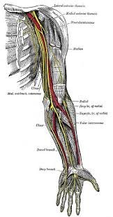
Things to identify
Median Nerve
Crucial for diagnosing carpal tunnel syndrome and performing median nerve blocks.
Entrapment occurs beneath the flexor retinaculum at the wrist.
Ulnar Nerve
Assessed in cases of cubital tunnel syndrome and used in ulnar nerve blocks for hand procedures.
Radial Artery
Common site for arterial cannulation and arterial blood gas sampling.
Ulnar Artery
Important for assessing collateral hand circulation before radial artery procedures (e.g., Allen’s test).
Flexor Tendons of the Wrist
Evaluated in trauma and tenosynovitis; tendon glide studies help assess injuries and post-op function.
Palmaris Longus Tendon
Often used as a landmark for locating the median nerve and harvested for grafts in tendon repair.
Brachial Artery (distal, at elbow)
Landmark for distal nerve blocks, and an alternate site for blood pressure monitoring.
Olecranon Process
Key surface landmark for joint aspiration, trauma evaluation, and elbow dislocation reduction.
Elbow Joint Effusion
Easily detected by ultrasound to assess for inflammatory or septic arthritis.
Carpal Bones (e.g., scaphoid)
Important in evaluating occult wrist fractures, especially scaphoid fractures in FOOSH injuries. -

Things to identify
1. Internal Jugular Vein (IJV) Essential for central venous access; serves as a landmark for surrounding structures.
2. Common Carotid Artery (CCA) Critical artery; must be avoided during procedures; landmark for neck blocks.
3. Sternocleidomastoid Muscle (SCM) Anatomical reference for locating the IJV and carotid artery; important for block orientation.
4. Thyroid Gland (lobes + isthmus) Assessed for nodules, goiter, inflammation; guide for fine-needle aspiration.
5. Trachea Landmark for airway management, tracheostomy, and cricothyrotomy.
6. Cricothyroid Membrane Key site for emergency airway access.
7. Esophagus Important structure to avoid during airway and neck interventions.
8. Vertebral Artery Must be identified to avoid injury during deep cervical blocks.
9. Stellate Ganglion Target for sympathetic blocks in pain syndromes (e.g., CRPS, vascular insufficiency).
10. Cervical Nerve Roots (e.g., C5–C7) Identified for selective nerve root blocks and diagnosing radiculopathy.
11. Brachial Plexus (interscalene region) Main target for regional blocks of the upper extremity.
12. Anterior Scalene Muscle Landmark muscle for interscalene brachial plexus block.
13. Middle Scalene Muscle Defines lateral boundary of the brachial plexus sheath.
14. Hypoechoic Lymph Nodes Evaluation of cervical lymphadenopathy in infection or malignancy.
15. Submandibular Gland Differentiated from lymph nodes and masses; relevant for sialolithiasis or infections. -

Superficial Structures
1. Ribs Reason: Bony landmarks for orientation; fractures or adjacent pathology often seen here.
2. Intercostal Muscles Reason: Important for detecting trauma and guiding needle placement safely between ribs.
3. Pectoralis Major Muscle Reason: Superficial guide for chest wall vascular access and mass evaluation.
4. Serratus Anterior Muscle Reason: Landmark and target for serratus plane blocks to manage thoracic pain.
5. Internal Mammary (Thoracic) Vessels Reason: Critical to avoid during parasternal procedures like biopsy or drain placement.
Deep Structures
6. Pleura (Parietal and Visceral Pleurae) Reason: Key to diagnosing pneumothorax (absence of sliding) and pleural effusion.
7. Lung Surface (A-lines, B-lines) Reason: A-lines show normal lung aeration; B-lines point to interstitial diseases or pulmonary edema.
8. Subclavian Vessels (Vein and Artery) Reason: Essential for ultrasound-guided central line placement and recognizing vascular injury risks.
9. Diaphragm (Thoracic Side) Reason: Check for diaphragmatic movement disorders or injury; vital for thoracoabdominal procedures.
10. Pericardium (Anterior Chest View) Reason: Allows identification of pericardial effusion or tamponade, especially during trauma evaluations.
